

DiO (DiOC18(3)) 细胞膜绿色荧光探针
分享
收藏
 推荐应用
推荐应用
产品介绍
DiO是一种亲脂性的荧光染料,可以用来染细胞膜和其它脂溶性生物结构。进入细胞膜后,DiO在整个细胞膜上扩散,最佳浓度时可以使整个细胞膜染色。DiO在进入细胞膜之前荧光非常弱,当与细胞膜结合后其荧光强度大大增强,被激发后可以发出绿色的荧光,具有很高的淬灭常数和激发态寿命。可以用标准的FITC滤光片检测。
DiO通常不会明显影响细胞的生存力,因此被广泛用于正向或逆向的,活的或固定的神经等细胞或组织的示踪剂或长期示踪剂(long-term tracer)。除了最简单的细胞膜荧光标记外,还可以用于检测细胞的融合和粘附,检测发育或移植过程中细胞迁移,通过FRAP(Fluorescence Recovery After Photobleaching)检测脂在细胞膜上的扩散,检测细胞毒性和标记脂蛋白等。
产品特色
| 同义名(Synonym) | DiOC18(3); DiO perchlorate; 3,3-Dioctadecyloxacarbocyanine perchlorate |
| CAS 号(CAS NO.) | 34215-57-1 |
| 分子式(Molecular Fomular) | C53H85ClN2O6 |
| 分子量(Molecular Weight) | 881.72 |
| 外观(Appearance) | 橘色至红色固体 |
| Ex/Em | 484/501 nm |
| 推荐滤光器(Filter) | XF23-Omega, 31001-Chroma |
| 纯度(Purity) | >97% |
| 溶解性(Solubility) | 溶于DMF,DMSO,乙醇 |
| 结构式(Structure) | 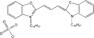 |
存储条件
冰袋运输。产品-20 ºC干燥避光保存,有效期1年。
COA
已发表文献
相关产品
联系我们